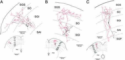
Fig. 2.
Camera lucida drawings of GAD67-GFP-positive neurons that were filled with biocytin during whole-cell patch-clamp recordings. The axons project mainly dorsally to the SGS (A and B) or to both SGS and SGP (C). Red lines indicate axon collaterals; black, the cell soma and dendrites. SO, optic layer; SAI, stratum album intermedium or intermediate white layer; SAP, stratum album profundum or deep white layer; PAG, periaqueductal gray layer.

