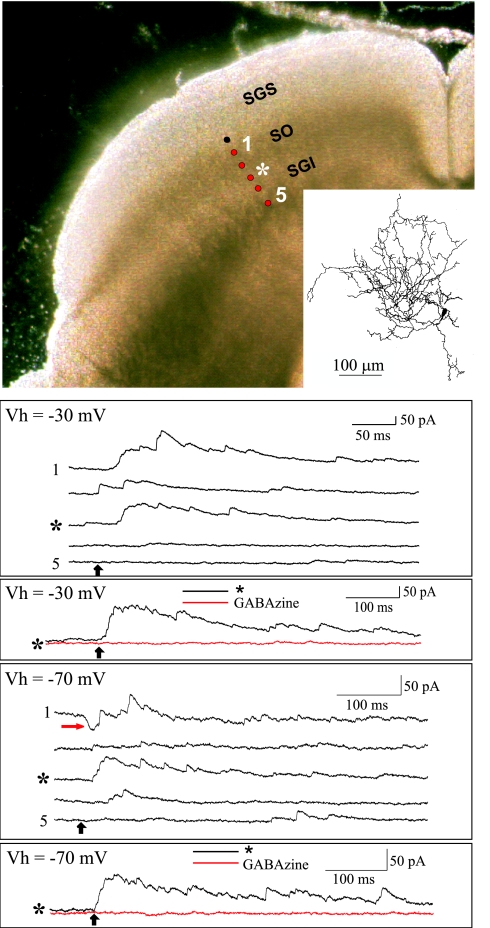
Fig. 3.
Examples of synaptic currents evoked in SGS cells by photostimulation in SGI. (Upper Left) The photomicrograph is of a coronal slice through the superior colliculus and shows the location of the patch-clamped cell (black dot) and the photostimulation sites (red dots). Sites 1–5 were 100–500 μm from the recorded cell, respectively. (Inset) Camera lucida drawing of the patch-clamped cell. (Top traces) Outward currents evoked by photostimulation at each of the sites indicated by the red dots while the cell was clamped at −30 mV. (Middle traces) Currents evoked by stimulation at site 3 (*), which was 300 μm from the recorded cell and in SGI, were blocked by gabazine. (Bottom five traces) Currents evoked while the cell was clamped at −70 mV. Both EPSCs (red arrow) and IPSCs were evoked when the photostimulation site was in the optic layer (site 1), but the predominant responses were IPSCs when the stimulus was located in SGI (site 3, *). (Bottom trace) Currents evoked by stimulation at site 3 (*) while the cell was clamped at −70 mV. These outward currents were blocked by the addition of gabazine (red trace), but few if any EPSCs were unmasked.

