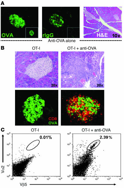
Figure 2. Anti-OVA IgG drives OT-I cell infiltration into the pancreas.
(A) Anti-OVA IgG binds to the islets of Langerhans without causing acute inflammation. Staining for OVA (left), bound anti-OVA IgG (by detecting rIgG; middle), and H&E (right) in the pancreata of RIP-mOVA mice 24 hours after treatment with 1,000 μg anti-OVA IgG or 1,000 μg control rIgG (insets). Transferred anti-OVA IgG bound to islet cells (middle) but did not trigger infiltrative lesions (right). (B and C) Diabetes results from OT-I cell infiltration into the pancreas. H&E, CD8 (red), and OVA (green) staining on pancreatic sections (B) and flow cytometric analysis of total live pancreatic cells (C) from RIP-mOVA mice 7 days after transfer of either OT-I cells alone or together with 1,000 μg anti-OVA IgG. Vα2 and Vβ5 indicate the presence of OT-I TCR+ cells. Destructive cellular infiltrates were observed after the transfer of T cells and anti-OVA IgG, including massive intraislet CD8+ OT-I cell accumulation, whereas T cells transferred alone did not accumulate in the pancreas nor induced inflammatory responses.

