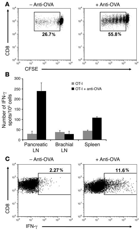
Figure 3. Anti-OVA IgG enhances functional antigen presentation in the pancreatic lymph node.
(A) OT-I cell proliferative responses. Pancreatic lymph node cells isolated from RIP-mOVA mice 3 days after transfer of CFSE-labeled OT-I cells and anti-OVA IgG. Dot plots are gated on CD8+CFSE+ cells. Proliferation was enhanced 2-fold in recipients of OT-I cells and anti-OVA IgG. (B and C) OT-I cell effector differentiation. ELISPOT analysis of IFN-γ production by total lymph node and splenic cells (B) and intracellular IFN-γ staining of pancreatic lymph node OT-I cells (C) isolated from RIP-mOVA mice 5 days after treatment. Bars show mean ± SD for triplicate wells of 1 representative experiment out of 3. Dot plots are gated on CD8+Vα2+Vβ5+ cells. Differentiation of OT-I cells into IFN-γ–producing effectors was significantly (P = 1.06 × 10⁻⁶) enhanced in the presence of anti-OVA IgG.

