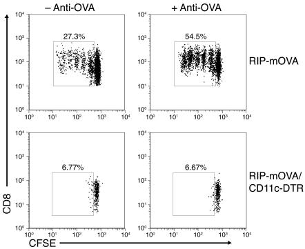
Figure 5. DCs are required for steady-state and antibody-enhanced OT-I cell proliferation in the pancreatic lymph node.
Flow cytometric analysis of pancreatic lymph node cells isolated from DT-treated RIP-mOVA and RIP-mOVA/CD11c-DTR mice 3 days after transfer of CFSE-labeled OT-I cells alone (left panels) or together with anti-OVA IgG (right panels). Dot plots are gated on CD8+CFSE+ cells and represent 1 of 3 independent experiments. OT-I cell proliferative responses were abolished in the absence of DCs (bottom panels).

