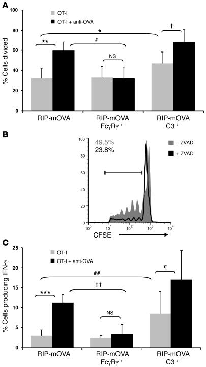
Figure 7. Antibody-enhanced OT-I cell proliferation and effector cell differentiation is dependent on activating Fcγ receptors.
(A and B) OT-I cell proliferative responses in WT, FcγRγ–/–, and C3–/– mice. Flow cytometric analysis of pancreatic lymph node cells isolated from RIP-mOVA, RIP-mOVA FcγRγ–/–, and RIP-mOVA C3–/– mice (A) and ZVAD-treated and untreated RIP-mOVA C3–/– mice (B) 3 days after transfer of CFSE-labeled OT-I cells and anti-OVA IgG. The percentage of cells divided (A) and the numbers in the histogram (B) were calculated from FACS plots gated on CD8+CFSE+ cells found after the undivided peak. Bars show mean ± SD for at least 5 mice per treatment group. *P = 0.033; †0.0085; **P = 0.00040; #P = 0.00060; NS: P = 0.46. Histogram represents 1 of 2 independent experiments. Activating FcγRs were required for antibody-enhanced proliferation, whereas C3 negatively regulated T cell proliferation in a ZVAD-inhibitable manner. (C) OT-I cell effector differentiation in WT, FcγRγ–/–, and C3–/– mice. Intracellular IFN-γ staining of pancreatic lymph node cells isolated from mice 5 days after treatment. The percentage of cells producing IFN-γ was calculated from IFN-γ–positive cells among CD8+Vα2+Vβ5+-gated populations. Bars show mean ± SD for at least 3 mice per group. ***P = 1.06 × 10⁻⁶; ††P = 0.00048; ##P = 0.016; ζP = 0.037; NS: P = 0.28. Antibody enhancement of IFN-γ production was intact in C3–/– but not FcγRγ–/– mice.

