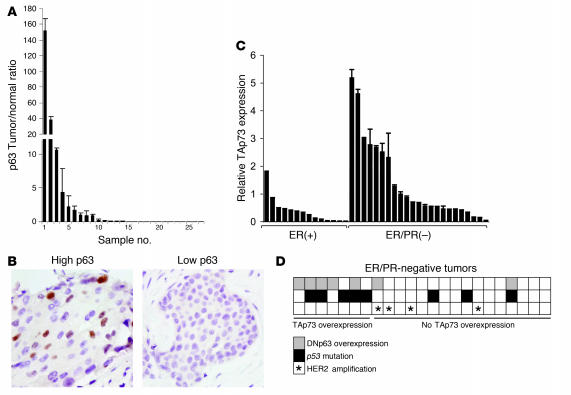
Figure 1. Coexpression of ΔNp63 and TAp73 in triple-negative primary breast tumors.
(A) Overexpression of p63 in primary microdissected invasive breast carcinomas relative to specimen-matched normal luminal epithelia. The ratio of tumor/normal p63 mRNA was determined by QRT-PCR. (B) Nuclear p63 protein correlated with p63 mRNA expression, as assessed by immunohistochemistry of representative tumors from A. Photomicrographs demonstrate low and high expression of p63 mRNA. Original magnification, ×100. (C) TAp73 was overexpressed in ER/PR-negative tumors. Shown is QRT-PCR for TAp73 in 14 ER-positive and 23 ER/PR-negative primary breast carcinomas. Statistical significance was analyzed using both a mean-value approach (ER-positive, 0.373 ± 0.126; ER/PR-negative, 1.381 ± 0.303; P = 0.0172, 2-way Student’s t test) and a binning approach whereby tumors exhibiting p73 expression more than 2-fold the mean of the sample set were categorized as high and the rest as low (P = 0.0309, Fisher’s exact test). (D) Expression of TAp73 correlated with ΔNp63 overexpression (P = 0.0107, Fisher’s exact test) and with p53 mutation (P = 0.0257, Fisher’s exact test) in ER/PR-negative primary breast carcinomas. Levels of ΔNp63 were determined by QRT-PCR, and p53 mutation was determined by cDNA sequencing. Note that TAp73/ΔNp63 coexpression was not observed in Her2-overexpressing tumors as assessed by QRT-PCR.

