
Figure 2.
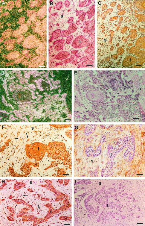
Colocalization of MMP-13, MT1-MMP, and MMP-2 expression in vulvar SCCs. A: Dark-field exposure of an in situ hybridization showing MMP-13 mRNA positive tumor cells in a primary vulvar SCC (patient 7). B: Bright-field photomicrograph corresponding to panel A. C: Immunostaining of a section parallel to that shown in panel B with anti-MT1-MMP antibody, showing MT1-MMP-positive tumor cells and stromal fibroblasts. D: Dark-field exposure showing expression of MMP-13 mRNA by tumor cells in a recurrent vulvar SCC (patient 9). E: Bright-field exposure corresponding to panel D. F: Immunostaining of a section parallel to that shown in panel E with anti-MT1-MMP antibody showing MT1-MMP-positive tumor cells and stromal fibroblasts (arrows). G: Immunostaining of a section parallel to that shown in panel E with anti-MMP-2 antibody showing MMP-2 expression in stromal cells and in tumor cells in the periphery of tumor islands. H: Immunostaining of a section parallel to that shown in panel E with anti-MMP-13 antibody showing MMP-13-positive tumor cells and stromal fibroblasts (arrows). I: Control immunostaining of section parallel to that shown in panel E using preimmune mouse ascites fluid in place of primary antibody shows no positive staining. t, tumor; s, stroma. Bars: 24 μm (A-E, I) and 10 μm (F-H).