
Figure 3.
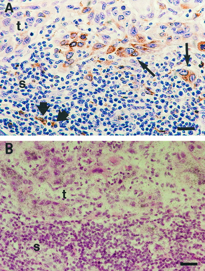
Colocalization of MMP-13- and MMP-9-expressing cells in vulvar SCCs. A: Immunostaining of a vulvar SCC (patient 3) with anti-MMP-9 antibody, showing MMP-9-positive tumor cells (thin arrows) and mononuclear inflammatory cells (thick arrows). B: In situ hybridization of a section parallel to that shown in A with MMP-13 antisense probe showing MMP-13 expression by tumor cells. Bars: 6 μm.