
Figure 4.
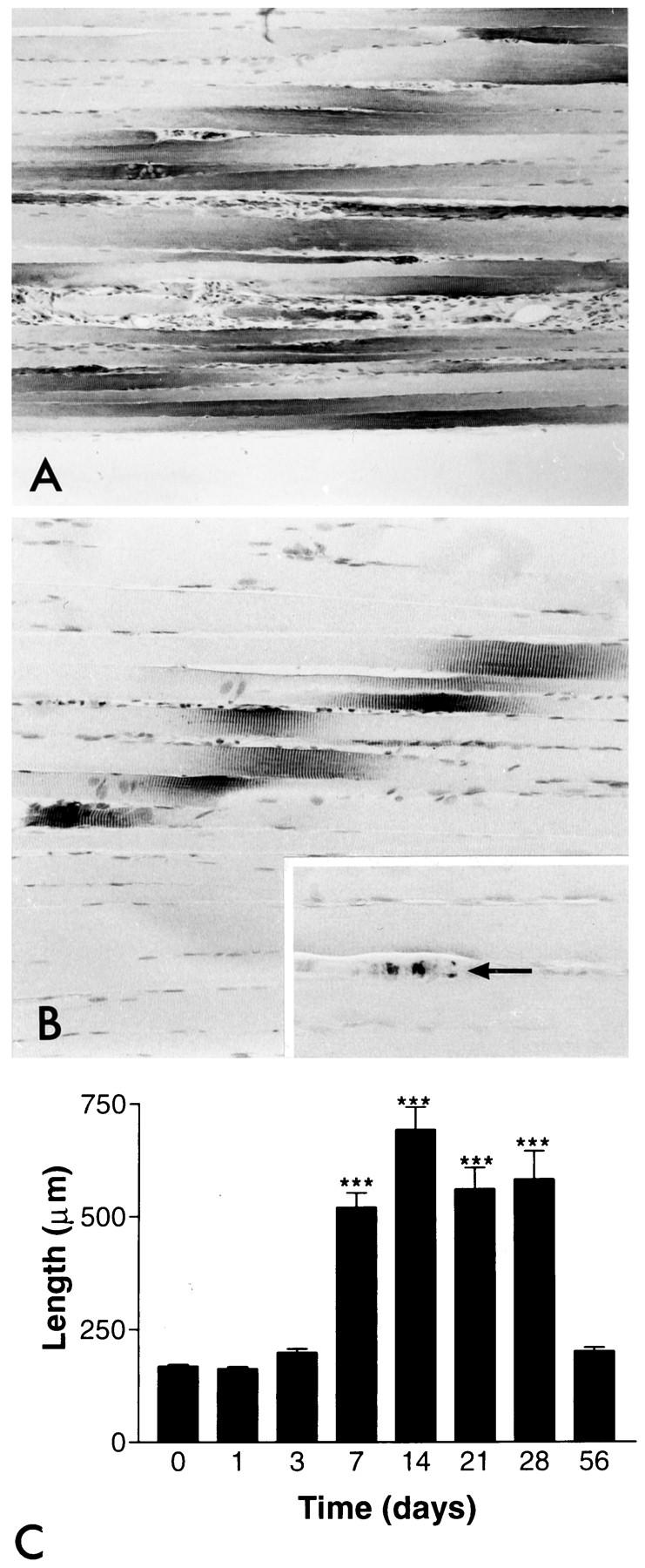
The pattern and intensity of nestin immunoreactivity after denervation and re-innervation. A: Nestin immunoreactivity 28 days after denervation. The length of immunoreactivity has clearly increased. Note that the denervated myofibers have become atrophic. B: Restored nestin distribution after complete re-innervation 56 days after denervation. The length of the nestin immunoreactivity has returned to the same as in intact myofibers (cf Figure 1A ▶ ). Inset: Regenerated axons have also become detectable with neurofilament immunostaining (arrow). Magnification, ×79 (A), ×158 (B), and ×247 (inset). C: The length of nestin immunoreactivity in the NMJ regions on each group is shown as a time course after denervation. Numbers are mean values ± SEM; ***P < 0.001.