
Figure 3.
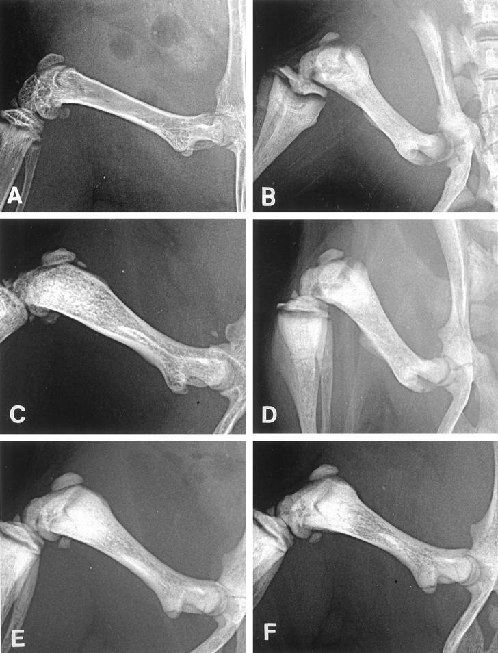
X-ray findings of femurs in a young wild-type mouse (A), a young op/op mouse (B), an aged op/op mouse (C), and a GM-CSF-treated (D), an IL-3-treated (E), and a GM-CSF- plus IL-3-treated (F) young op/op mouse. A: The femur of the wild-type mouse has a wide marrow cavity and clearly distinguishable cortical bones. B: The femur of the young op/op mouse is short and thick, associated with diffuse bone density in the medullary cavity and indistinguishable cortical bones. C: In the aged op/op mouse, the medullary cavity is formed in the femur, is distinguished from the bone cortex, and shows a lacy pattern of bone density. D to F: In the treated op/op mice, the bone marrow cavity is formed, and the cortical bones are distinguished from the cavity. Slight lacy patterns of density are found in the medullary cavity.