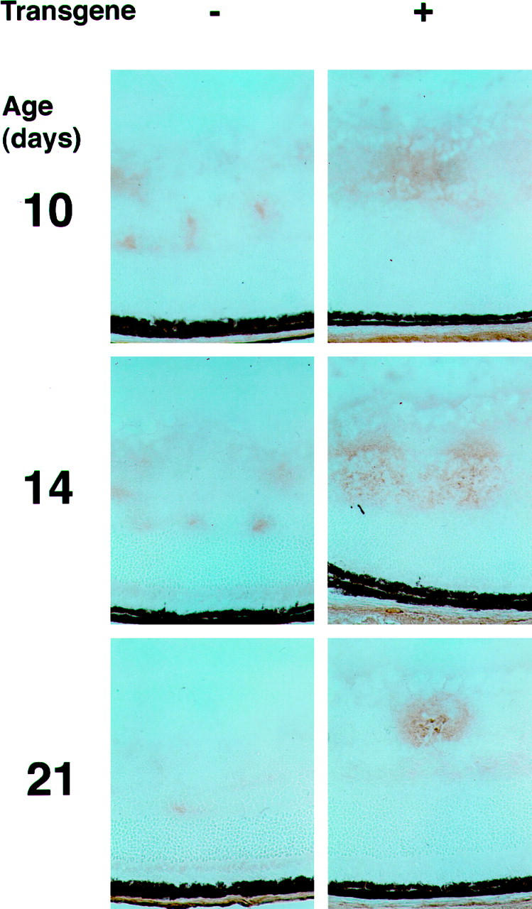
Figure 10.

Immunohistochemistry for FGF2 in the retinas of P10, P12, and P21 rho/PDGF-A transgene-positive and -negative mice with ischemic retinopathy. rho/PDGF-A+/− mice crossed with wild-type mice and resultant litters were placed in 75% oxygen at P7 and removed to room air at P12. Mice were sacrificed at P10, after 3 days of hyperoxia; P14, 2 days after removal from hyperoxia to the relative hypoxia of room air; and P21, 9 days after onset of the hypoxic period. Eyes were rapidly removed, and frozen sections were immunohistochemically stained for FGF2. At each time point, there was increased staining for FGF2 in transgene-positive mice compared with that in transgene-negative mice. The FGF2 staining occurred in the inner part of the retina, where both the ectopic astrocytes and retinal blood vessels were located.
