
Figure 4.
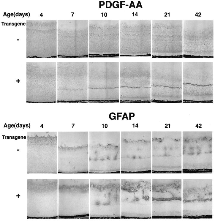
Time course of immunohistochemical staining for PDGF-AA and GFAP. Transgene-positive and -negative mice from the rho/PDGF-A3 line were sacrificed on P4, P7, P10, P14, P21, and P42. The eyes were frozen, sectioned, and stained for PDGF-AA or GFAP as described in Materials and Methods. Expression of PDGF-A is seen in the photoreceptors of transgene-positive mice starting on P7 and shows prominent staining, particularly in photoreceptor terminals, at all subsequent time points. At P4 and P7 in both transgene-positive and -negative mice, there is staining for GFAP in the nerve fiber layer, the normal location for astrocytes. At P10 and later time points, there is GFAP staining in the nerve fiber layer and in columns that extend into the inner nuclear layer, which likely represents perivascular astrocytes. The staining in both locations appears greater in transgene-positive mice and at P21 and P42, and there is a continuous horizontal streak of GFAP-positive cells that is not present in littermate controls.