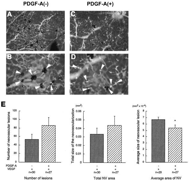
Figure 7.
Transgenic mice that express both PDGF-A and VEGF do not have less NV than mice that express only VEGF. rho/PDGF-A+/− mice were crossed with rho/VEGF+/+ mice. At P21, the pups were perfused with fluorescein-labeled dextran, and retinal whole mounts were examined by fluorescence microscopy. Mice were genotyped after quantitation of NV. rho/PDGF-A transgene-negative mice (A: ×100; B: ×400) had numerous areas of NV surrounded by retinal pigmented epithelial cells typical of rho/VEGF mice. rho/PDGF-A transgene-positive mice (C: ×100; D: ×400) also showed numerous areas of NV that appeared smaller with less participation of retinal pigmented epithelial cells. E: The number of neovascular lesions, the area per lesion, and the total area of NV per retina were measured by image analysis. There was no significant difference in the number of lesions or total area of NV per retina, but the average area of lesions was significantly less in mice that expressed both PDGF-A and VEGF, compared with mice that expressed only VEGF (*P = 0.0364 by two-tailed t-test).

