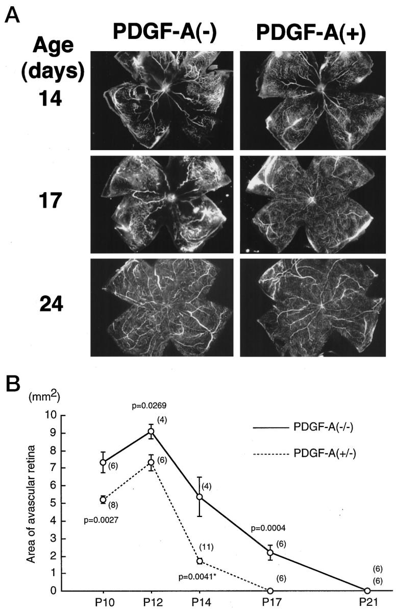
Figure 8.

rho/PDGF-A transgene-positive (PDGF-A+) mice with ischemic retinopathy have less retinal vascular nonperfusion than littermate controls (PDGF-A−). Rho/PDGF-A+/− mice were crossed with wild-type mice, and resultant litters were placed in 75% oxygen at P7 and removed to room air at P12. At P10, P12, P14, P17, P21, and P24, mice were perfused with fluorescein-labeled dextran, and retinal whole mounts were prepared. The retinas were examined by fluorescence microscopy, and the area of vascular nonperfusion per retina was measured by image analysis. The mice were genotyped at the time of sacrifice, but the examiner was masked for genotype. A: At P14, PDGF-A− mice showed large areas of vascular nonperfusion, which are typical for wild-type mice after 5 days of hyperoxia, but PDGF-A+ mice showed only a small area of nonperfusion. At P17, PDGF-A+ mice had complete revascularization of the retina, but PDGF-A− mice still have a large area of nonperfusion. At P24, both types of mice were completely revascularized. B: The mean (± SEM) area of avascular retina was plotted for each time point for PDGF-A− and PDGF-A+ mice. The number of retinas used to calculate the mean for each time point (shown in parenthesis adjacent to each point) was determined by the litter sizes and chance breakdown of genotypes. At each time point, PDGF-A+ mice had significantly less avascular retina by two-tailed t-test than PDGF-A− mice.
