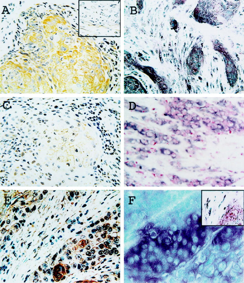
Figure 1.

Expression of u-PA, u-PAR, and PAI-2 in esophageal SCC. Immunostaining with antibodies to u-PA (A), u-PAR (C), and PAI-2 (E). Representative data of in situ hybridization using antisense probes for u-PA (B), u-PAR (D), and PAI-2 (F). Positive staining was observed in cancer cells (A–F) and the fibroblasts surrounding them (B, E, insets in A and F).
