
Figure 1.
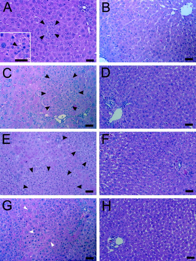
Emergence and expansion of small hepatocyte-like cells after PH in retrorsine-exposed rats. A, C, E, and G: Retrorsine-exposed rat livers at 3, 7, 10, and 14 days post-PH, respectively (H&E). B, D, F, and H: Control rat livers at 3, 7, 10, and 14 days post-PH, respectively (H&E). Aggregates of small hepatocyte-like cells are seen in easily identifiable clusters of 3 to 6 cells by 3 days post-PH in retrorsine-exposed rats (A), approach lobule size by 7 days post-PH (C), and coalesce into large patches by 10 days post-PH (E). Small hepatocyte-like cells occupy nearly 50% of the parenchyma by 14 days post-PH (G). No small hepatocyte-like cells are observed in control/PH rats at any time point (B, D, F, H). Bar, 50 μm. Black arrows indicate small hepatocyte-like cells; white arrows indicate megalocytes. Inset in A shows a higher magnification of a different field.