
Figure 2.
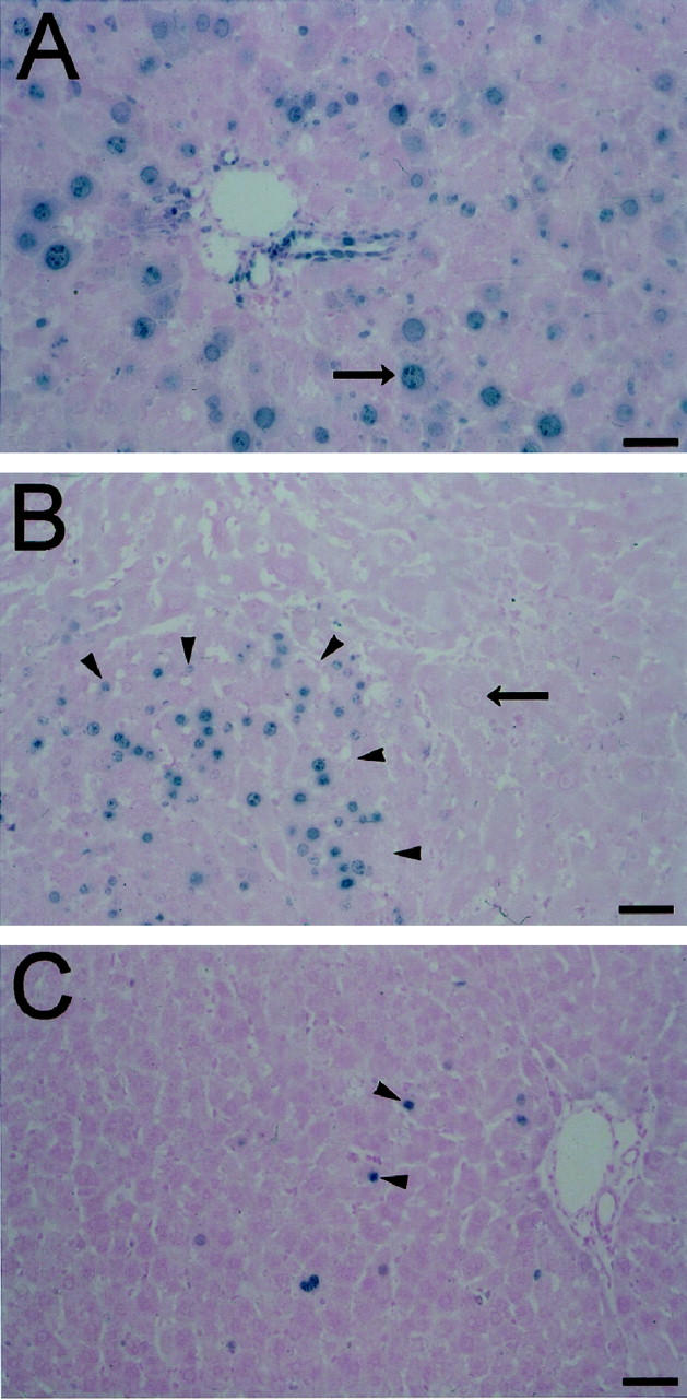
Indirect immunoperoxidase detection of Ki-67 nuclear antigen in retrorsine-exposed rat livers after PH. A: Retrorsine-exposed rat liver section labeled with the nuclear antigen Ki-67 3 days after PH. B: Retrorsine-exposed rat liver section labeled with the nuclear antigen Ki-67 14 days after PH. C: Retrorsine-exposed rat liver section labeled with the nuclear antigen Ki-67 30 days after PH. At 3 days post-PH (A) hepatocyte megalocytosis is clearly visible. The majority of hepatocytes and bile duct epithelial cells stain positive for nuclear antigen Ki-67 (blue chromagen), indicating an attempt at replication, although hepatocytes do not complete the cell cycle. By 14 days post-PH (B), only small hepatocyte-like cells show evidence of proliferation, and only when the liver mass was fully reconstituted at 30 days post-PH (C) did the proliferative activity of the small hepatocyte-like cells diminish. Bar, 50 μm. Long arrows indicate megalocytes and short arrows indicate small hepatocyte-like cells.