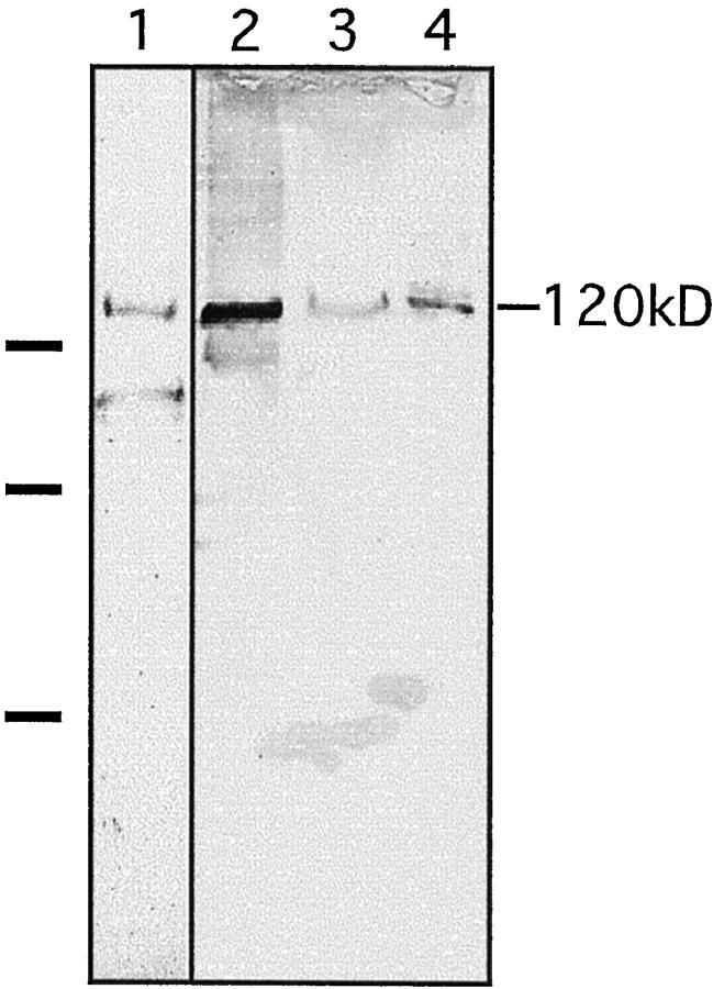
Figure 2.
Identification of the 120-kd soluble ectodomain of collagen XVII in amniotic fluid and pemphigoid blister fluid. Immunoblotting of the shed ectodomain extracted from the epidermis (lane 1) and from keratinocyte culture medium (lane 2). Antibodies used for the blot were anti-NC16a in lane 1 and Ecto1 in lane 2. The soluble ectodomain was immunoprecipitated with the anti-NC16a antibody from amniotic fluid (lane 3) and bullous pemphigoid blister fluid (lane 4) and immunoblotted with the Ecto1 antibody. On the left, the migration positions of molecular mass markers are indicated: from top to bottom 112, 80, and 50 kd. The lower band of approximately 90 kd in lane 1 represents a degradation product of the shed ectodomain. 1

