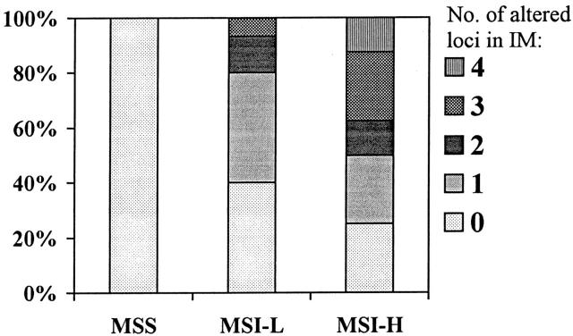
Figure 3.
Frequency of altered microsatellites in IM of patients with coexisting MSI-H, MSI-L, and MSS tumors. The number of altered microsatellite markers in IM (0, 1, 2, 3, and 4) of different tumor groups is indicated by different filling patterns, which are defined on the right side of the Figure ▶ . IM samples from patients with MSI-H tumors had significantly more loci altered than those from MSI-L or MSS (P = 0.003).

