
Figure 4.
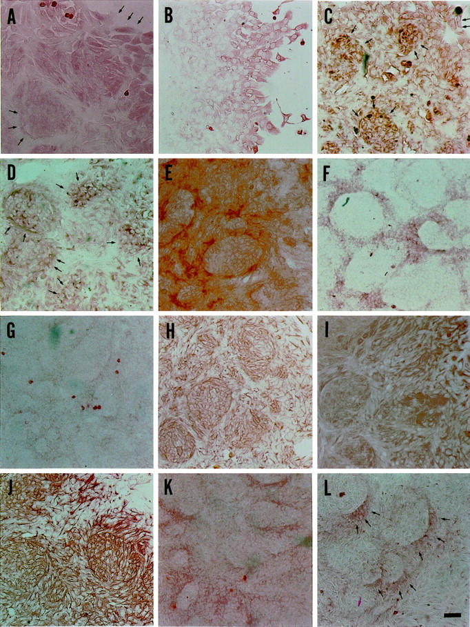
Immunocytochemical analysis of duct-like structure-emerged colonies on Day 8 to 10. Albumin (A), transferrin (C), and AAT (D) are present in the cells at the periphery of colonies and in those surrounded by duct-like structures. AAT is detectable in the cells at the periphery of colonies as colonial spots. AFP (B) is expressed only in the cells at the periphery of colonies after Day 9. GGT (E), CK19 (F), CK14 (G), and OC.10 (L) are present in the cells constituting duct-like structures, although CK14 expression is faint. CK7 (H), CK18 (I), OV6 (J), and BD.2 (K) are expressed in all cells covering the duct-like structure-emerged colonies. Cells at the periphery of colonies express OV6 more strongly than those in the central parts of colonies. Original magnifications, ×100. Arrows indicate positive cells. Scale bar, L, 50 μm.