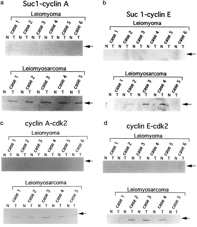
Figure 4.
Levels of cdk-associated cyclin A and cyclin E and levels of cdk2 associated with cyclin A or cyclin E. Lysates prepared from surgically resected tissues (200 μg) were precipitated with p13suc1-Sepharose (for cyclin A and cyclin E blotting) or immunoprecipitated with anti-cyclin A or anti-cyclin E antibodies (for cdk2 blotting). The precipitates were subjected to further immunoblotting analysis with the respective antibodies. Suc 1-cyclin A (a), Suc 1-cyclin E (b): Precipitation by p13suc1-Sepharose followed by cyclin A or cyclin E blotting, respectively. Cyclin A-cdk2 (c), Cyclin E-cdk2 (d): Immunoprecipitation by anti-cyclin A or anti-cyclin E antibodies followed by cdk2-blotting, respectively. N, normal tissue; T, tumor tissue.

