
Figure 2.
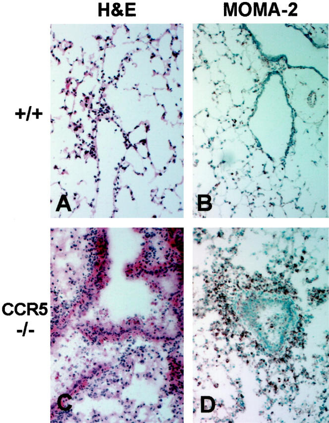
Inflammatory response to influenza A in lungs of control and CCR5-deficient mice. All histology sections are from lung tissues taken at day 2 postinfection with 5 HAU of influenza A; original magnification, ×100. A and B: Sections from +/+ lungs show H&E staining of only a few inflammatory cells characteristic for this early time point (A) and MOMA-2 staining of a few, sparse resident alveolar macrophages (B). C and D: Sections from CCR5−/− lungs show H&E staining of massive inflammatory cell infiltration and interstitial pneumonitis (C) and intense MOMA-2 staining of the infiltrating macrophages (D).