
Figure 3.
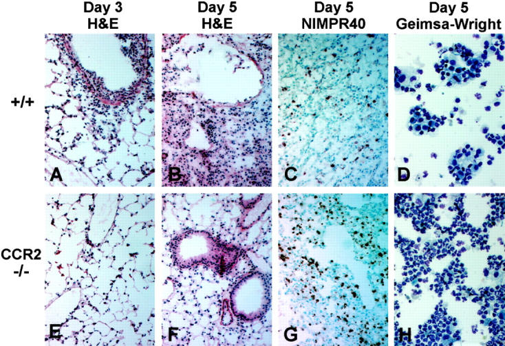
Inflammatory response to influenza A in lungs of control and CCR2-deficient mice. All histology sections are from lung tissue taken at days 3 and 5 postinfection with 5 HAU of influenza A; original magnification, ×100. Cytological preparations are BAL samples taken at day 5 postinfection; original magnification, ×400. A–C: Sections from +/+ lungs show H&E staining of the acute inflammatory cell infiltrate typically seen 3 days postinfection (A), the areas of chronic inflammation and tissue consolidation, thickening of the septal walls, and disorganization of the bronchial epithelial cells typically seen 5 days postinfection (B), and NIMPR40 staining of the relatively few neutrophils seen at this time point in +/+ mice (C). D: Giemsa-Wright staining of the BAL cells obtained from the +/+ mice 5 days postinfection. E–G: Sections from CCR2−/− lungs show H&E staining of less cellular infiltration 3 days postinfection (E), absence of the epithelial cell damage and consolidating pneumonitis that is seen in control mice at day 5 postinfection (F), and relatively intense NIMPR40 staining of the accumulating neutrophils, which is the dominant leukocyte population present in CCR2−/− lungs 5 days postinfection (G) . H: Giemsa-Wright staining of CCR2−/− BAL cells obtained at day 5 postinfection.