
Figure 3.
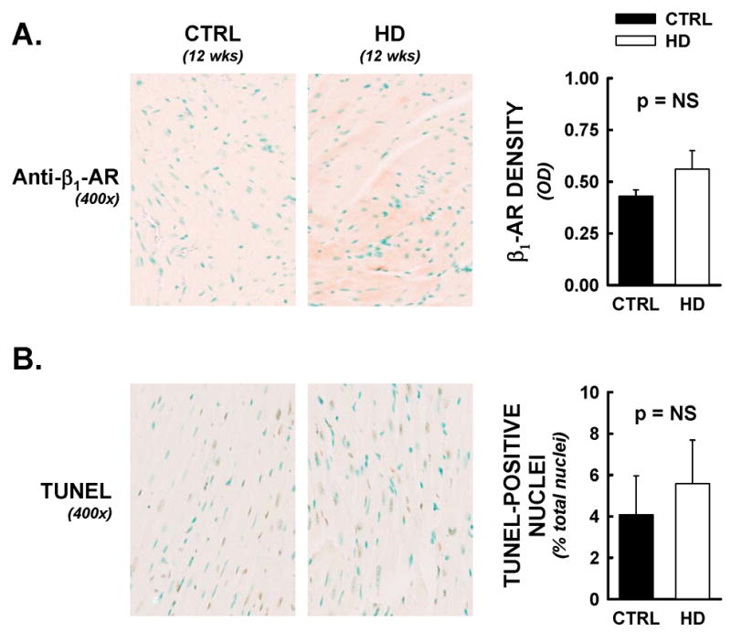
Cardiac β1-adrenergic receptor densities and myocyte apoptotic events were not altered in HD mice. Following functional analyses, hearts were assessed for β1-adrenergic receptor densities and DNA nicks by immunohistochemical methods. All data are from animals sacrificed at 12 weeks of age. Panel A) Representative photomicrographs from anti- β1 adrenergic receptor labelling (400x). No significant differences were observed between HD hearts and littermate controls, by optical density analysis. Panel B) TUNEL staining was employed as an indirect marker of apoptotic events. TUNEL positive nuclei (brown nuclei) were counted by an automated digital imaging approach, and expressed as a percentage of DNAse positive control (maximal nuclear staining) from serial sections. Percentages of total nuclei that were TUNEL-positive were not significantly different between treatment groups.