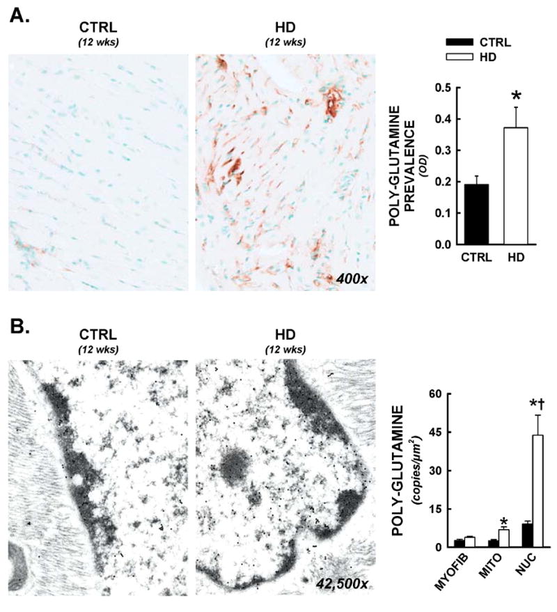
Figure 4.

Nuclear and mitochondrial residence of mutant huntingtin in R6/2 mice. Following functional analyses, cardiac tissues were prepared for both light microscopy and electron microscopy immunohistochemistry. All data are from animals sacrificed at 12 weeks of age. Panel A) Representative photomicrographs from anti-polyglutamine staining (antibody raised against poly-glutamine repeats >37, 400x). Increased prevalence of the expanded polyglutamine sequence was visualized in both cytosolic and nuclear spaces. Total cardiac polyglutamine prevalence was significantly elevated in HD hearts by optical density analysis. Panel B) Immunogold electron microscopy for intracellular distributions of mutant huntingtin. Immunohistochemistry was conducted as above, with a gold-colloid secondary antibody (10nm particles, visualized as electron dense circles under tunneling electron microscopic examination). Images of myocytes with longitudinal orientation were captured and nuclear, mitochondrial, and myofibrillar areas were delineated and calibrated, then positive copies for the expanded polyglutamine sequence were counted, and expressed per nuclear, mitochondrial, or myofibrillar area. Mean data is represented in histogram format. *, p<0.05 versus littermate control.
