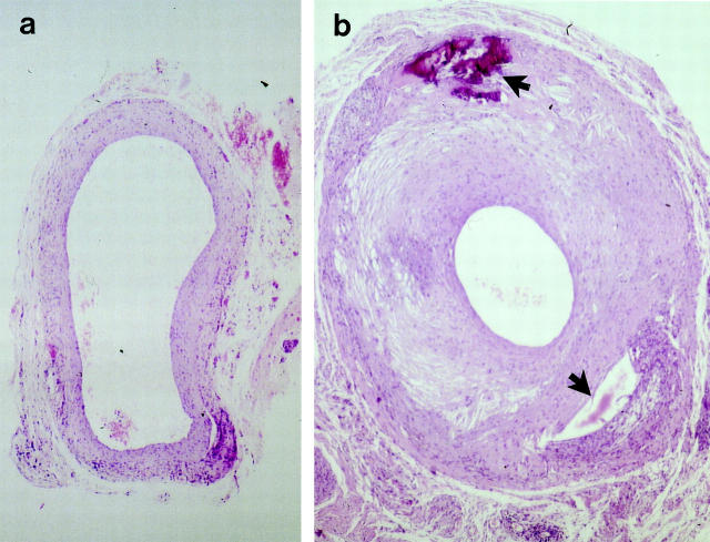
Figure 1.
Hematoxylin-eosin (HE)-stained sections of mouse vein grafts. Under anesthesia, vena cava veins of apoE+/+ (a) or apoE−/− (b) mice were removed and isografted into carotid arteries of apoE +/+ or apoE−/−, respectively. Animals were sacrificed 8 weeks after surgery, and the grafted tissue fragments were fixed in 4% phosphate-buffered formaldehyde, pH 7.2, embedded in paraffin, sectioned, and stained with HE. Upper arrow indicates necrosis, and lower arrow indicates loss of tissues possibly due to necrosis. Original magnification, ×40.

