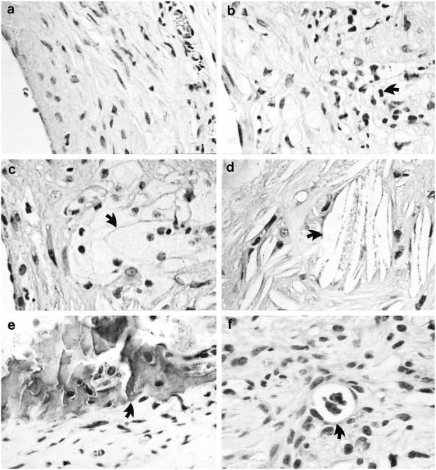
Figure 3.

Morphological characteristics of vein graft atheroma. HE-stained sections of 8-week neointima (a) of vein grafts from apoE+/+ mice and vein graft atheroma (b-f) from apoE−/− mice. Arrows indicate typical examples of infiltrated mononuclear cell (b), foam cell (c), cholesterol crystal (d), calcified necrotic core (e), and neovasculature (f).
