
Figure 4.
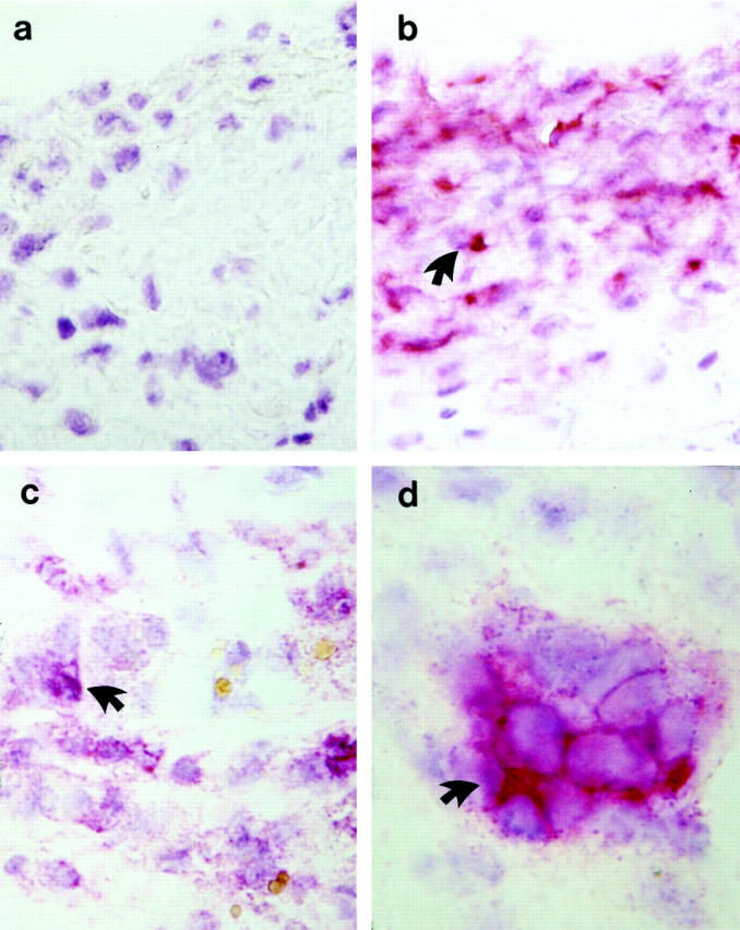
Immunohistochemical staining demonstrates the presence of α-actin-, MAC-1-, and CD4-positive cells in vein grafts. Sections derived from vein grafts at 8 weeks were labeled with normal rat serum (a), a mouse monoclonal antibody against α-actin conjugated with alkaline phosphatase (b), rat monoclonal antibodies against MAC-1-positive leukocytes (CD11b/18; c) or against CD+ T-cells (d) and developed with alkaline phosphatase-anti-alkaline phosphatase techniques. A counterstaining (blue) with hematoxylin was performed. Arrows indicate examples of typical positive cells (red). Original magnifications, ×400 (a-c) and ×1000 (d).