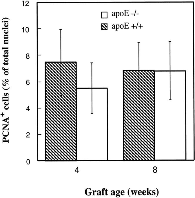
Figure 5.
Immunofluorescent labeling for PCNA-positive cells in vein grafts. Sections derived from vein grafts of apoE+/+ and apoE−/− mice at 4 and 8 weeks were labeled with a rabbit anti-PCNA antibody and visualized by swine anti-rabbit Ig conjugated with FITC. Cell nuclei were stained with Hoechst 33258 for 1 minute. Positive cells and total nuclei were counted in a 400× field. Two fields of each section were enumerated, and 3 sections per vein graft were selected. Data are means ± SD of 4 animals per time point in each group.

