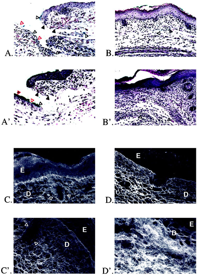
Figure 1.

H&E staining and confocal scanning laser microscopy of gestation day 16 and 19 fetal rat skin wounds. A: Gestation day 16 excisional skin wound, 12 hours after injury. Original magnification: ×200. The wound edge (solid black triangles) and wound base (open red triangles) are indicated. There is minimal inflammatory infiltrate. Note the presence of blue vital dye used to mark the location of the fetal wound (open black triangles). B: Gestation day 16 excisional skin wound, 72 hours after injury. Original magnification: ×100. There is complete healing, with restoration of normal dermal architecture and regeneration of hair follicles. Small areas containing blue vital dye are visible in the dermis (open black triangles). A′: Gestation day 19 excisional skin wound, 12 hours after injury. Original magnification: ×200. The wound edge (solid black triangles) and wound base (open red triangles) are indicated. There is moderate inflammatory infiltrate. Note again the presence of blue vital dye in the wound (open black triangles). B′: Gestation day 19 excisional skin wound, 72 hours after injury. Original magnification: ×100. To the right of the wound is a small area of nonwounded skin with intact hair follicles. There is complete epithelialization over the wound, but the normal dermal architecture has been disrupted by unorganized collagen deposition with no regeneration of hair follicles. Small areas of blue vital dye are also visible in the dermis (open black triangles). C: Confocal microscopy of nonwounded E19 fetal skin. Original magnification: ×630. The epidermis (E), dermis (D), and a portion of a hair follicle (open white triangles) are indicated. D: Confocal microscopy of an E16 embryo excisional wound, 72 hours after injury. Original magnification: ×630. In addition to hair follicle regeneration (open white triangles), collagen architecture is indistinguishable from nonwounded gestation day 19 skin (E16 + 72 hours postinjury = E19). C′: Confocal microscopy of nonwounded N1 fetal skin. Original magnification: ×630. The epidermis (E), dermis (D), and a portion of a hair follicle (white triangles) are again indicated. D′: Confocal microscopy of an E19 embryo excisional wound, 72 hours after injury. Original magnification: approximately ×630. There is no hair follicle regeneration, and the collagen architecture is disorganized and denser when compared to nonwounded neonatal day 1 controls (E19 + 72 hours postwounding = N1).
