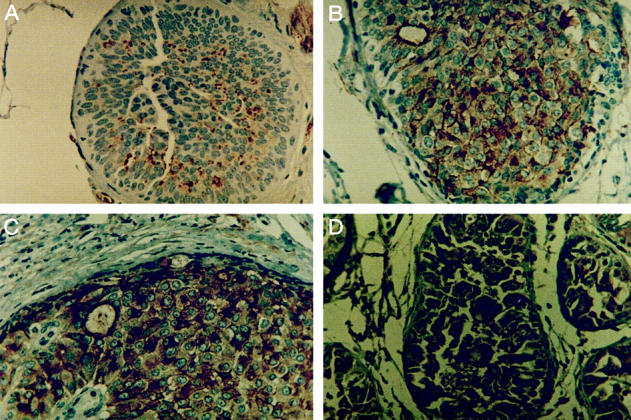
Figure 1.

Immunohistochemical analysis of clusterin expression in proliferative lesions and DCIS. A: Clusterin negative ductal hyperplasia without atypias. B: Atypical ductal hyperplasia showing positive staining. C and D: Strong cytoplasmic clusterin staining is seen in a low-grade DCIS with intermediate nuclear grade and absence of necrosis (C) and in a high-grade DCIS (D). All fields are magnified ×400. All sections were counterstained with hematoxylin.
