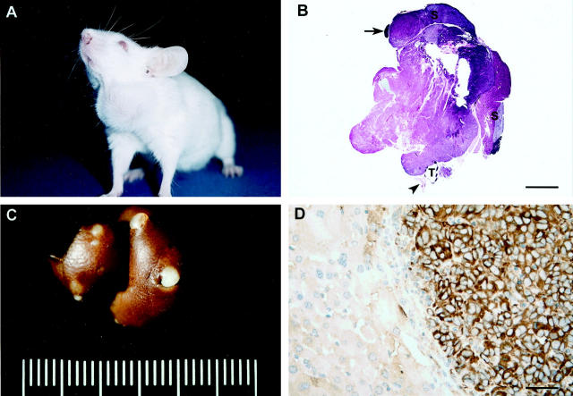
Figure 4.
Metastatic ATC in a ret/PTC1+ p53−/−mouse (170 days old). A: Note the asymmetrical swelling in the ventral cervical region corresponding to the thyroid tumor. B: Bilateral ATC with complete penetration through the thyroid capsule (3+ invasion) and marked asymmetrical enlargement on the left. Adjacent salivary glands (S) and lymph node (arrow) are not affected. T, trachea; arrowhead, esophagus. HE staining. Bar, 4.8 mm C: Multiple metastases of ATC in the liver. D: Immunohistochemical stain for thyroglobulin in the liver. The metastatic thyroid tumor cells are labeled positively for thyroglobulin, but the adjacent hepatocytes are negative. Results of immunohistochemical staining for ret were similar (not shown). Hematoxylin counterstain. Bar, 80 μm.

