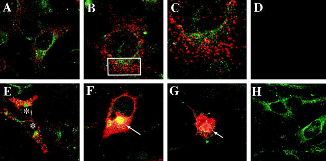
Figure 2.
Confocal analysis of α-synuclein expression in transfected GT1-7 cells. Cells were double-immunolabeled with antibodies against murine α-synuclein (red) and the neuronal marker microtubule-associated protein 2 (green) and imaged with the laser-scanning confocal microscope. A: Control nontransfected cells showed mild punctuate α-synuclein immunostaining associated with the cytoplasm and some neuritic processes. B: ST cells showed intense immunoreactivity associated with granular cytoplasmic structures. C: Detail at higher magnification of the rectangular area presented in (B). D: Negative control with the primary antibodies inactivated. E, F, and G: ST cells showed intense immunoreactivity associated with granular cytoplasmic structures (*) and dense intracellular inclusions (arrows). H: AST cells showed microtubule-associated protein 2 immunoreactivity but minimal or none α-synuclein immunostaining.

