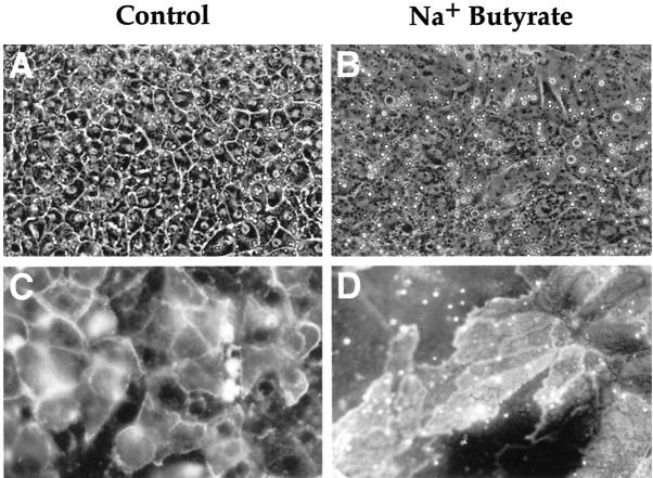
Figure 9.
Morphology of E12 hepatoblasts cultured in vitro and expression of HES6 and BDS7. Phase-contrast (A and B, ×120) and immunofluorescence (C and D, ×320) microscopy of hepatoblasts from E12 liver cultured with standard medium (A and C) and in the presence of Na+ butyrate (B and D). C and D: Immunodetection of HES6 (C) and BDS7 (D).

