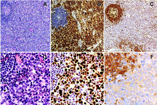
Figure 2.

Lack of SHP-1 expression by a transformed large-cell cutaneous T cell lymphoma involving lymph node. The images represent ×100 low-power (A–C) and ×400 high-power (D–F) magnifications. Methylene blue was used as a counterstain. A and D: H&E staining. B: Anti-CD3 antigen staining (T-cell receptor-associated antigen). C: Anti-CD79a staining (immunoglobulin-associated B cell antigen). E: Anti-mib1 (Ki67) staining (antigen expressed in cycling but not resting cells). F: anti-SHP-1 staining. Note a residual nonmalignant, partially involuted follicle with preserved to mildly expanded mantle zone in the upper left corners of all photographs.
