
Figure 5.
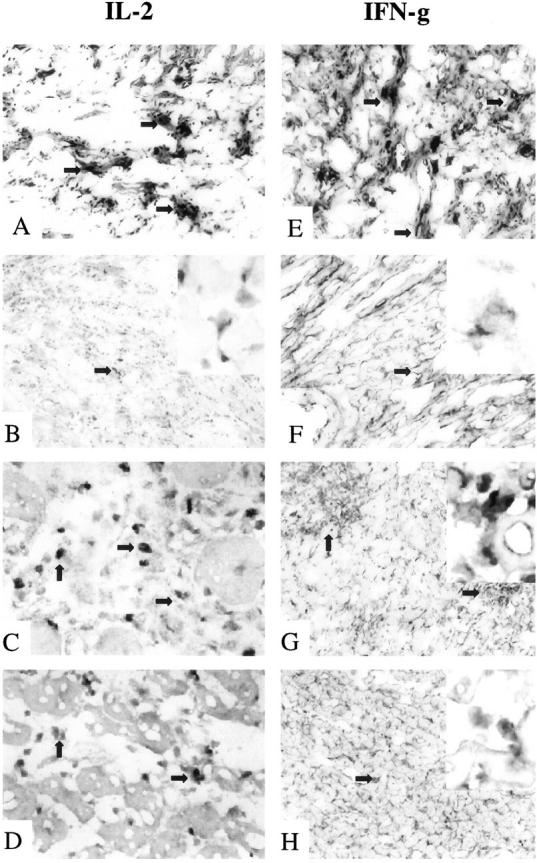
Immunohistochemical analysis of IL-2 and IFN-γ expression in rat cardiac allografts. Examination of IL-2 (A−D) and IFN-γ (E−H) shows that the tolerant state in RIB-5/2 mAb-treated hosts at 120 days was accompanied by basal staining for Th1 cytokines (B and F). In contrast, recipients undergoing chronic rejection showed dense expression of both IL-2 and IFN-γ (A and E). Adoptively transferred recipients undergoing rejection of test cardiac allografts after infusion of CD4-depleting mAb (OX36) showed moderate levels of Th1 cytokines (C and G). This contrasted with only mild IL-2 and IFN-γ deposition (D and H) after adjunctive course of CS1 peptides (OX36 + CS1). Original magnifications, ×100 (A, B, E−H) and ×270 (C and D) (n = 2). Arrows indicate positive labeling, and enlargements of those areas are included in the upper right insets of B, F, G, and H (original magnifications, ×420).