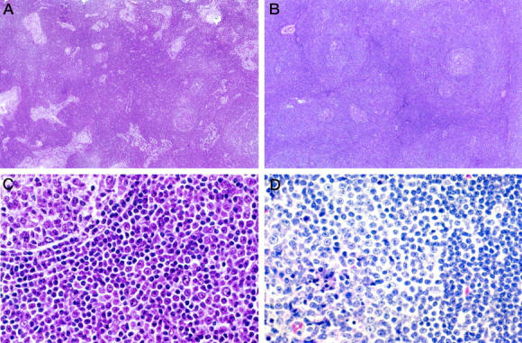
Figure 1.

Histological features of I-SLL. A: A low-power view of an I-SLL (case 4) that demonstrated extensive sinus preservation, reactive follicles, and only scattered pale proliferation centers. B: A low-power view of an I-SLL (case 9) with perifollicular proliferation centers around reactive follicles. C: A higher magnification of case 9 showing a reactive follicle (upper left) with its thin mantle zone surrounded by cells which closely resemble those of a proliferation center. D: A higher magnification of an I-SLL (case 13) that demonstrated reactive follicles partially colonized by proliferation center type I-SLL cells. The residual reactive follicular center cells are best seen in the lower left corner associated with tingible body macrophages.
