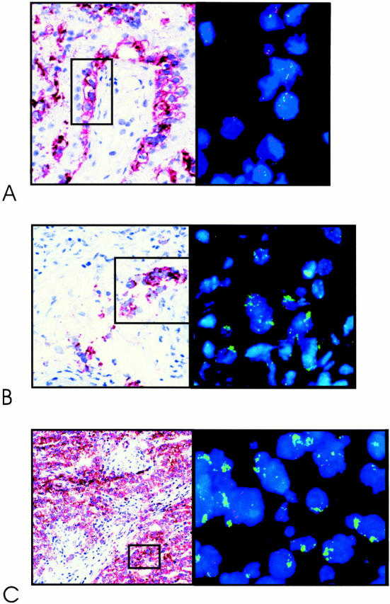
Figure 3.

Representative examples of double-fluorescent in situ hybridization on frozen tissue sections, 4-μm thick, using a probe specific for the centromeric region of chromosome 12 (red), and YAC#5 (green), mapped within the shortest region of overlap of amplification (see Figure 1 ▶ ). Shown are carcinoma in situ (A); microinvasive seminoma (B); and invasive seminoma (C) . The tumor cells are identified by the direct enzymatic alkaline phosphatase detection method (stained in red) on a parallel tissue section.
