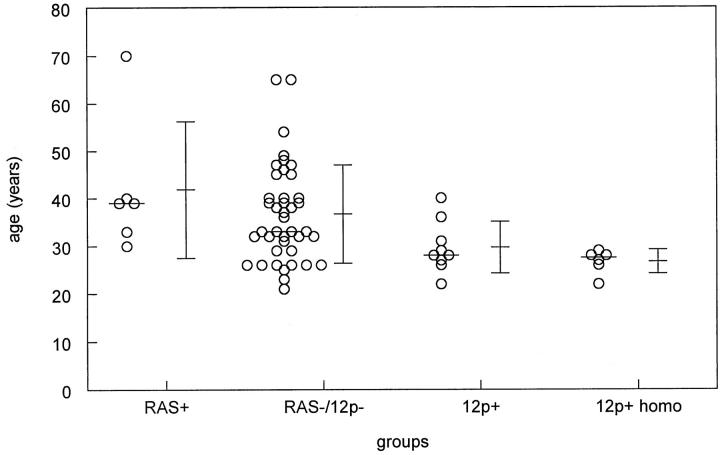
Figure 5.
Schematical representation of the ages of patients at clinical presentation with a seminoma with and without 12p amplification and RAS mutation (mean, average, and standard deviations are indicated). In addition, the ages of the subgroup of patients with a homogeneous 12p amplification are shown. No differences were found between the ages of patients with a seminoma without either of these aberrations and those with a RAS mutation. However, those with a 12p amplification-positive seminoma showed a borderline significant difference compared to the control group, whereas a significant difference was found in case only patients with a homogeneous 12p amplification were included (P = 0.023). Abbreviations: RAS+, RAS mutation; RAS−/12p−, no RAS mutation or 12p amplification; 12p+, 12p amplification both heterogeneously and homogeneously present; 12p+ homo, homogeneous 12p amplification.

