
Figure 5.
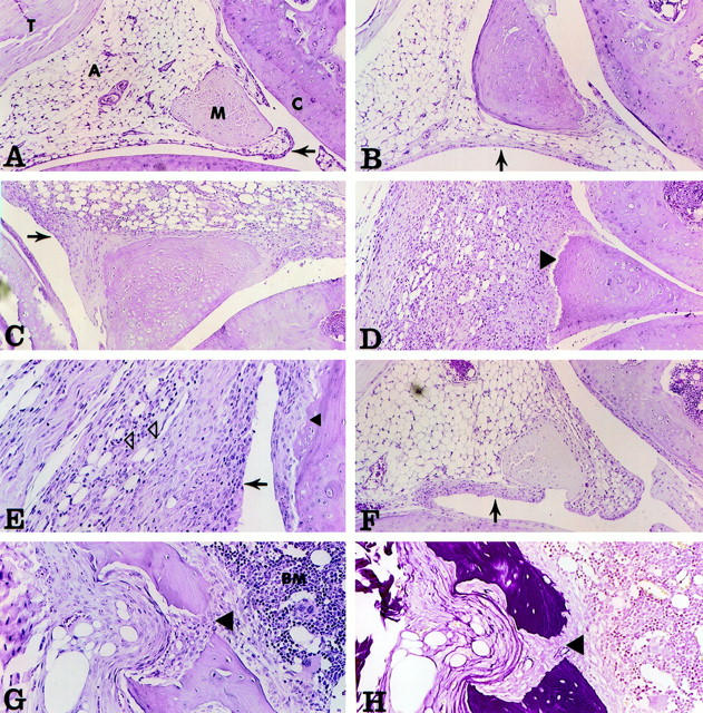
Histopathology of mouse knee joints. Adenovirus vectors were injected into the knee joints of mice, and the animals were sacrificed at various times following administration. Knee joints were fixed, decalcified, paraffin-embedded, and stained using H&E and EVG techniques. Tissues were assessed using pathological criteria as shown in Table 1 ▶ . Original magnification: ×100 (A–D and F); ×200 (E, G, and H). A: Contralateral joint of AdmuOSM, day 7. A, Adipocytes; M, meniscus; C, articular cartilage; T, tendon. Note the normal appearance and thickness of the synovial lining (arrow). B: Addl70, day 7. The synovial lining shows little if any thickening (arrow). C: AdmuOSM, day 4. Note the substantial increase in the thickness of the synovial lining (arrow). D: AdmuOSM, day 7. The synovial lining is extensively thickened and the entire synovium is infiltrated with mononuclear and some polymorphonuclear cells (PMN), with a concomitant decrease in adipocyte presence. Note the eroded miniscal outer surface (arrow). E: AdmuOSM, day 7. Higher magnification shows infiltration of the synovium, the PMN being clearly visible (open arrowheads). Note also the erosion of the articular cartilage adjacent to synovial tissue (closed arrowhead). F: AdmIL-6, day 7. The synovial lining shows a minor increase in thickness (arrow). G and H: AdmuOSM, day 7, showing matching fields stained with H&E and EVG, respectively. The synovial tissue has eroded the articular cartilage and underlying bone, and has penetrated the bone marrow (BM) (large arrowhead).