
Figure 1.
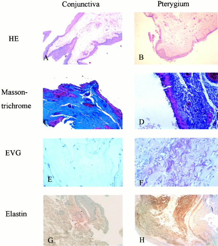
Histological studies of pterygia. A: H&E stain of pterygia showing prominent subepithelial connective tissue in the pinguecular part of pterygia (original magnification, ×100). B: H&E stain of normal conjunctiva (original magnification, ×100). C: Masson’s trichrome stain of the pinguecular part in pterygia shows strong staining of collagen in the subepithelial connective tissue (original magnification, ×400). D: Masson’s trichrome stain of normal conjunctiva reveals weak staining of collagen in the subepithelial connective tissue (original magnification, ×400). E: Verhoeff’s elastic stain of the pinguecular part in pterygia reveals prominent coarse elastic fibers in the subepithelial layer (original magnification, ×400). F: Verhoeff’s elastic stain of normal conjunctiva shows no elastic fibers (original magnification, ×200). G: Immunohistochemical staining of elastin in the pinguecular part of pterygium is positive in deeper subepithelial connective tissue (original magnification, ×100). H: Immunohistochemical staining of elastin is negative in normal conjunctiva (original magnification, ×100).