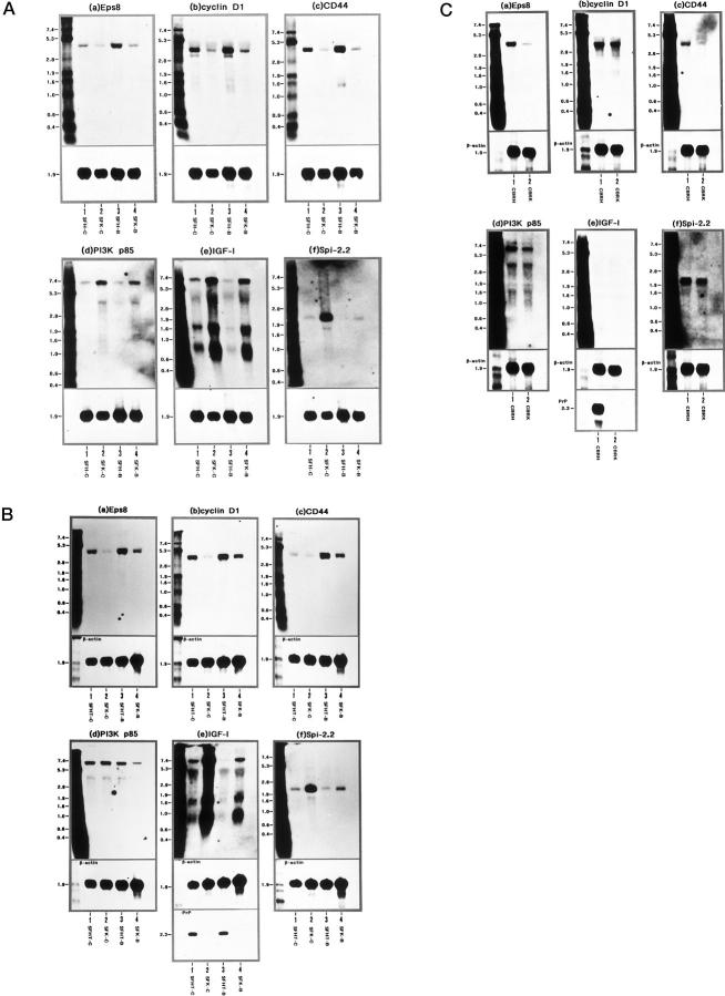
Figure 3.
Northern blot analysis of Eps8, cyclin D1, CD44, PI3K p85, IGF-I, and Spi-2.2 mRNA expression in SFK, SFH, and SFHT cells and brain tissues isolated from the PrP−/− and PrP+/+ mice. A distinct skin fibroblast cell line, designated SFHT, was established from the PrP+/− mice. SFHT cells were incubated for 72 hours in LS medium with (SFHT-B) or without (SFHT-C) inclusion of 50 ng/ml of bFGF for the last 24 hours before processing for RNA preparation. Total RNA was extracted from SFH-C, SFHT-C, SFK-C, SFH-B, SFHT-B, or SFK-B cells, and from the whole cerebral cortices isolated from the PrP−/− mice (CBRK) or from the PrP+/+ mice (CBRH). It was processed for Northern blot analysis by hybridization with digoxigenin (DIG)-labeled DNA probes specific for the Eps8 (a), cyclin D1 (b), CD44 (c), PI3K p85 (d), IGF-I (e), or Spi-2.2 (f) gene (upper panels), followed by rehybridization with the probe specific for the β-actin gene for standardization (lower panels) and, in limited experiments, with the probe specific for the PrP gene (lowest panels). A: Lanes 1–4 represent 2 μg of total RNA isolated from SFH-C (lane 1), SFK-C (lane 2), SFH-B (lane 3), and SFK-B (lane 4) cells, all of which were maintained for 4 months in vitro before starting the experiments. B: Lanes 1–4 represent 2 μg of total RNA isolated from the following cells: SFHT-C (lane 1), SFK-C (lane 2), SFHT-B (lane 3), and SFK-B (lane 4). SFHT cells were maintained for 4 months, whereas SFK cells were maintained for 7 months in vitro before starting the experiments. The lanes in C represent 6 μg of total RNA isolated from the brain tissues CBRH (lane 1) and CBRK (lane 2). The RNA size marker (kb) is shown in the left lane.

