
Figure 2.
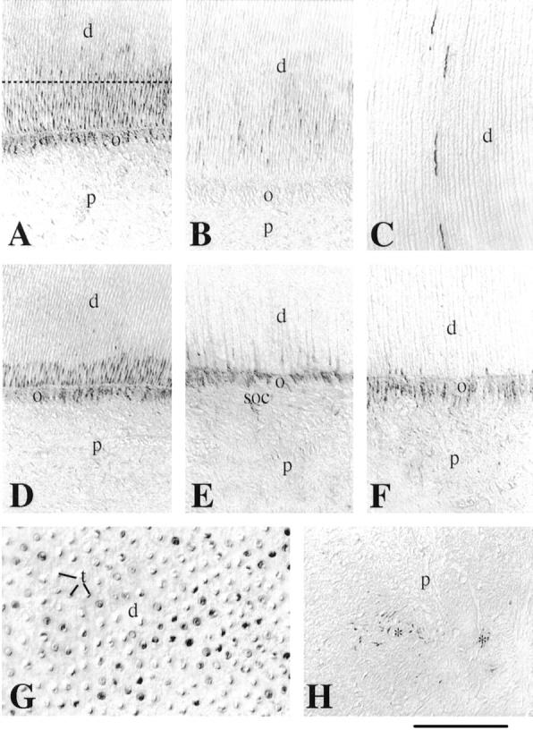
Immunohistochemical localization of nestin in sections of developing permanent human teeth. A: Crown area. Nestin immunoreactivity is observed in the cell bodies and processes of the odontoblasts. Note that the other pulp cells are negative for nestin. B: In the cusp of the crown region nestin staining is seen only in the odontoblastic processes. C: Nestin immunoreactivity is observed in some odontoblast processes near the dentin-enamel junction. D–F: From the middle of the crown (D) until the apical part of the root (F), nestin staining decreases in the odontoblastic processes and is limited in the odontoblastic cell bodies at the apical root area (F). Note the faint immunoreactivity in cells of the subodontoblastic layer at the root area (E). G: Section passing through the dotted line of A. Note the dentinal tubuli and the positive staining representing the odontoblastic processes. H: Immunoreactivity is observed in some pulp cells at the proximity of blood vessels (asterisks). d, dentin; o, odontoblasts; p, pulp; soc, subodontoblastic cells; t, dentinal tubuli. Scale bars, 50 μm (A, B, D–F, H) and 35 μm (C and G).