
Figure 1.
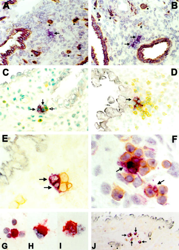
Immunohistochemistry of CD83+ and CD1a+ cells in early human pregnancy. Violet; VIP, brown; DAB, gray-blue; Vector blue, red; New fuchsin. A, C, and E: CD1a+ cells (violet, arrows); B, D, F, and J: CD83+ cells (violet, arrows). A: CD1a+ cells in decidua basalis exhibit a typical veiled appearance of DCs with long dendrites extending into the surrounding tissue. Brown: cytokeratin staining of invading FTBs (*) and endometrial glands (original magnification, ×160). B: Most CD83+ cells are located close to cytokeratin-positive endometrial glands (brown; original magnification, ×160). C: Typical location of CD1a+ cells in close vicinity to endometrial glands (gray-blue). CD1a+ cells are associated with a single CD3+ cell (brown, open head arrow; original magnification, ×250). D: CD83+ cells are often associated with CD3+ cells (brown) in clusters near an endometrial gland (gray-blue; original magnification, ×250). E: Association of CD1a+ cells with CD3+ cells (brown; original magnification, ×400). F: Association of CD83+ cells with CD3+ cells (brown; original magnification, ×630). J: CD83+ cells in a lymphatic vessel together with CD45+ lymphocytes (brown). * indicates invading FTBs indicating the tissue as decidua basalis (blue; original magnification, ×63). Cytocentrifuge preparations of enriched CD83+ cells demonstrate them to be often associated with lymphocytes (G; original magnification, ×630), to exhibit typical veiled morphology (H; ×1000) and to express HLA-DR (I; original magnification, ×1000).