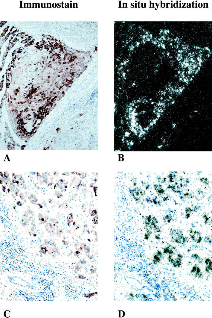
Figure 1.

A−D: Expression of COX-2 mRNA and immunoreactive-protein in malignant transitional cells in two patients (A and B, C and D) with grade III/high-grade histology. A and B: 100× bright-field and dark-field illumination of involved human bladder. A: COX-2 immunostaining showing brown reaction product, indicating COX-2-immuoreactive protein seen predominantly over infiltrating malignant transitional cells. No significant immunoreactivity is detected in surrounding stromal tissue. B: In situ hybridization using a COX-2-selective riboprobe. Black grains depict hybridization over submucosal infiltrating malignant transitional cells. Note that distribution of the COX-2 in situ hybridization signal in B is almost superimposable over the COX-2 immunoreactivity shown in A. C and D: 200× bright-field illumination of involved human bladder cancer from a patient with grade III/high-grade TCC. C: COX-2-immunoreactive protein is seen within islands of malignant epithelial cells. Surrounding stromal cells are COX-2-negative. D: Similar to the tissue section A and B; in situ hybridization shows COX-2 mRNA in cells that are COX-2 ir-protein-positive.
