
Fig. 2.
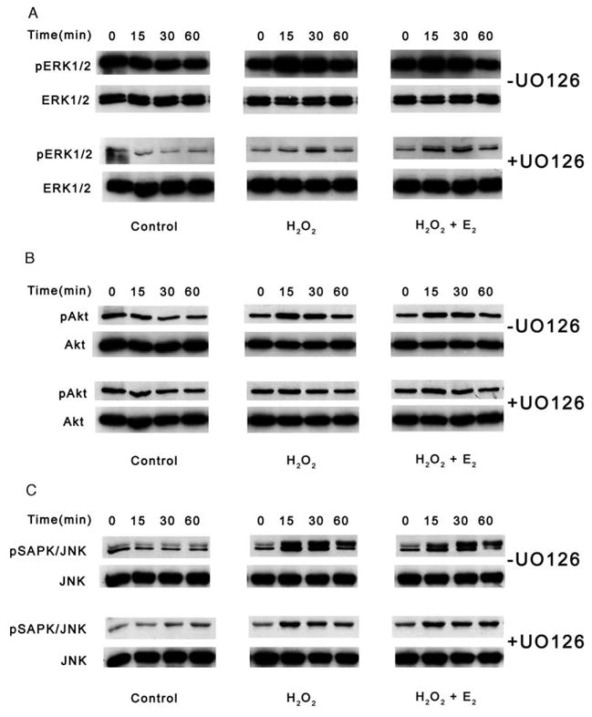
Western blot analyses of UO126 inhibition of MAPK signaling in HLE-B3 cells during an oxidative stress. Following a 1 h pretreatment with 10 μM UO126 (+UO126) or DMSO vehicle (−UO126), total cell lysates (20 μg protein/lane) were collected after 0, 15, 30 and 60 min of 100 μM H2O2±1 μM E2 exposure (middle and right columns) and analyzed for (A) ERK1/2, (B) Akt and (C) SAPK/JNK phosphorylation. Controls were not exposed to H2O2 or E2 (left column). This experiment was repeated twice with similar results.