
Fig. 5.
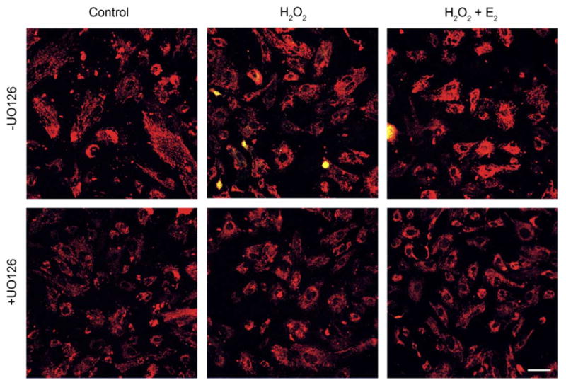
Confocal imaging of Δψm in BLECs exposed to H2O2 for 4 h. Following a 1 h pretreatment with 10 μM MEK1/2 inhibitor UO126 (+UO126) or DMSO vehicle (−UO126), cultures were treated with 100 μM H2O2±1 μM E2 (middle and right panels) and after 4 h stained with 5 μg/ml JC-1, a Δψm sensitive dye, for 30 min. Control cultures (left panels) received only UO126 or DMSO vehicle. No mitochondrial membrane depolarization was detected as no shift from red to green fluorescence occurred in H2O2-exposed cultures (middle panels). These images are typical of eight randomly chosen fields per treatment. Bar=20 μm.