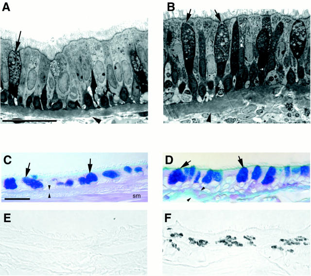
Figure 8.
Histopathological comparison of wall composition in the intrapulmonary bronchi of sensitized (B, D, and F) and nonsensitized (A, C, and E) rhesus monkeys. A and B: High resolution histopathological comparison of a control monkey (A) and an area of heavy inflammatory cell infiltration with epithelial hypertrophy and mucous goblet cell (arrows) hyperplasia of sensitized monkey (B) (scale bar, 20 μm). C and D: Differences in PAS/Alcian blue-positive mucous substance in the mucous goblet cells (arrows) of nonsensitized (C) and sensitized (D) rhesus monkeys (scale bar, 20 μm). E and F: Distribution of major basic protein in the epithelium of nonsensitized (E) and sensitized (F) rhesus monkeys (scale bar, 20 μm). Arrowheads indicate the basement membrane zone. Smooth muscle (SM).

