
Figure 4.
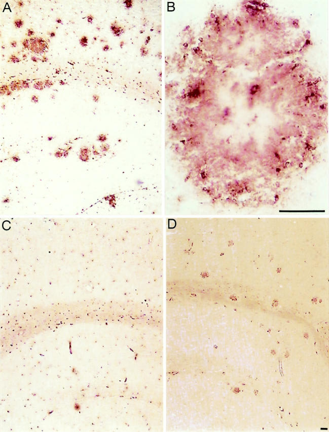
Activated microglia are capable to phagocytose. A: FA-11 (macrosialin) immunostaining of a section through the neocortex and hippocampus of a 23-month-old APP23 transgenic mouse shows strong macrosialin-positive amyloid plaque-associated microglia cells. B: A higher magnification of a single amyloid plaque visualizes macrosialin-positive microglia cell bodies that are surrounding the amyloid. Weaker labeling is found at the amyloid, thereby revealing the contours of the plaque. C: Microglia in the neocortex and hippocampus of a control mouse show a basal macrosialin staining in agreement with published data. 27 D: 2.4G2 staining of a section through the neocortex and hippocampus demonstrates an up-regulation of Fc receptors FcγRII and FcγRIII on activated microglia cells at amyloid plaques. Scale bar, 50 μm.